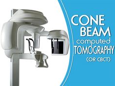
3D Images in Dentistry: Cone-Beam Computed Tomography
Who doesn’t love new technology? Whether it’s the latest smartphone or tablet, new innovations, gadgets and gizmos. Technology continues to push the envelope, turning mundane tasks into elegant procedures and solutions.
Dr. Jerry Vasilakos and the Planet Dentistry team in Toronto, Ontario feel the same way. You’ve perhaps already read about our love for the CAD/CAM milling machine.
Another of dental technology’s latest and greatest comes by way of Cone Beam Computed Tomography (CBCT). CBCT shows a 3D image of a patient’s jaw. This could be in its entirety, it could be focused on one particular area of the anatomy, or even the location of specific nerves and vessels. The scope is really quite incredible.
These 3D images are handy when trying to determine the quality of bone for implant placement. These cone beam images can give an extremely accurate representation of what exactly is in your jaw, in terms of thickness and depth.
And because these images are digital (thank you, technology!) there is software available that can actually place the implant onto the image, finding the optimal orientation so that we can maximize the amount of bone for the implant.
CBCT augments every stage of the dental practice, from treatment planning and diagnoses (like determining that there is even enough bone for an implant in the first place) to carrying out complex procedures (such as avoiding vital structures like nerves and vessels during the procedure).
Perhaps you’ve found what looks to be a troublesome growth in your mouth; if you’ve come in to Planet Dentistry, Dr. Jerry Vasilakos has access to a CBCT scanner which can reveal tumors or cysts that might be in the jaw.
Cone Beam Computed Tomography: it’s new, it’s exciting, and now available to those of us in the dental profession as never before.


